Loculated Pleural Effusion Ultrasound - Chest Radiograph - Radiography, ultrasound and chest ct reveal the presence of free or loculated pe, occasionally with images compatible with clots that may also reveal.
Loculated Pleural Effusion Ultrasound - Chest Radiograph - Radiography, ultrasound and chest ct reveal the presence of free or loculated pe, occasionally with images compatible with clots that may also reveal.. Occasionally you may see debris or loculations in the pleural effusion. The success rate is low when the effusion is loculated and septated. In healthy lungs, these membranes ensure that a small amount of liquid is present between the lungs. Pleural effusion symptoms include shortness of breath or trouble breathing, chest pain, cough, fever, or chills. It does tell you that it's going to be more difficult to do a thoracentesis, to actually.
Both the trocar and the modified seldinger techniques can be used. Technique for lung ultrasound in pleural effusion if the patient can sit forward. Pleural effusion, also called water on the lung, is an excessive buildup of fluid in the space between your lungs and chest cavity. Pleural effusions may result from pleural, parenchymal, or extrapulmonary disease. Pleural effusion symptoms include shortness of breath or trouble breathing, chest pain, cough, fever, or chills.
Peritoneal and meningeal relapse from lung adenocarcinoma ... from www.spandidos-publications.com Approximately 1 million people develop this abnormality each year in the empyema. If you have a patient with a loculated (or septated) pleural effusions are most often seen in exudative effusions and describe any effusion with fluid divided into pockets. Pleural effusion is an accumulation of fluid in the pleural cavity between the lining of the lungs and the thoracic cavity (i.e., the visceral and parietal pleurae). A role in selected clinical circumstances. Pleura l effusion seen in an ultra sound image as in one or more fixed pockets in the pleural space is said to be loculated pleural effusion.in. An ipc is sometimes more effective if the effusion is present on both sides of the chest (bilateral) or if there are large areas of localized fluid collections (loculated effusions). This is typically a chronic process. The patient should be comfortable, ideally sitting on the edge of the bed with arms folded forwards and.
Ultrasound signs of pleural effusions.
It is important to assess both the quantity of the pleural effusion and severity of the atelectasis. If you have a patient with a loculated (or septated) pleural effusions are most often seen in exudative effusions and describe any effusion with fluid divided into pockets. Pleural effusions may result from pleural, parenchymal, or extrapulmonary disease. A pleural effusion is accumulation of excessive fluid in the pleural space, the potential space that surrounds each lung. In this video briefly shown how we aspirate small amount of pleural fluid or loculated pleural effusion.for more videos please subscribe the channel.if you. Pleura l effusion seen in an ultra sound image as in one or more fixed pockets in the pleural space is said to be loculated pleural effusion.in. The lack of specificity is mainly due to the limitations of the imaging modality. The success rate is low when the effusion is loculated and septated. Pleural effusion (pleff), mostly caused by volume overload, congestive heart failure, and pleuropulmonary infection, is a common condition in critical care patients. There's always a small amount of liquid within this lining to help lubricate the. Diffuse nodules and opacification in right lung with compressive atelectasis. The patient should be comfortable, ideally sitting on the edge of the bed with arms folded forwards and. Ultrasound guided assessment of pleural effusion to determine and describe the size and site of the effusion.
Pleural effusion (pleff), mostly caused by volume overload, congestive heart failure, and pleuropulmonary infection, is a common condition in critical care patients. The loculated effusion located along the expected course of the fissure is well defined and elliptical, with pointed. Ct is also useful in the evaluation of loculated effusions, as seen in fig. Pleural effusion refers to a buildup of fluid in the space between the lungs and the chest cavity. Occasionally you may see debris or loculations in the pleural effusion.
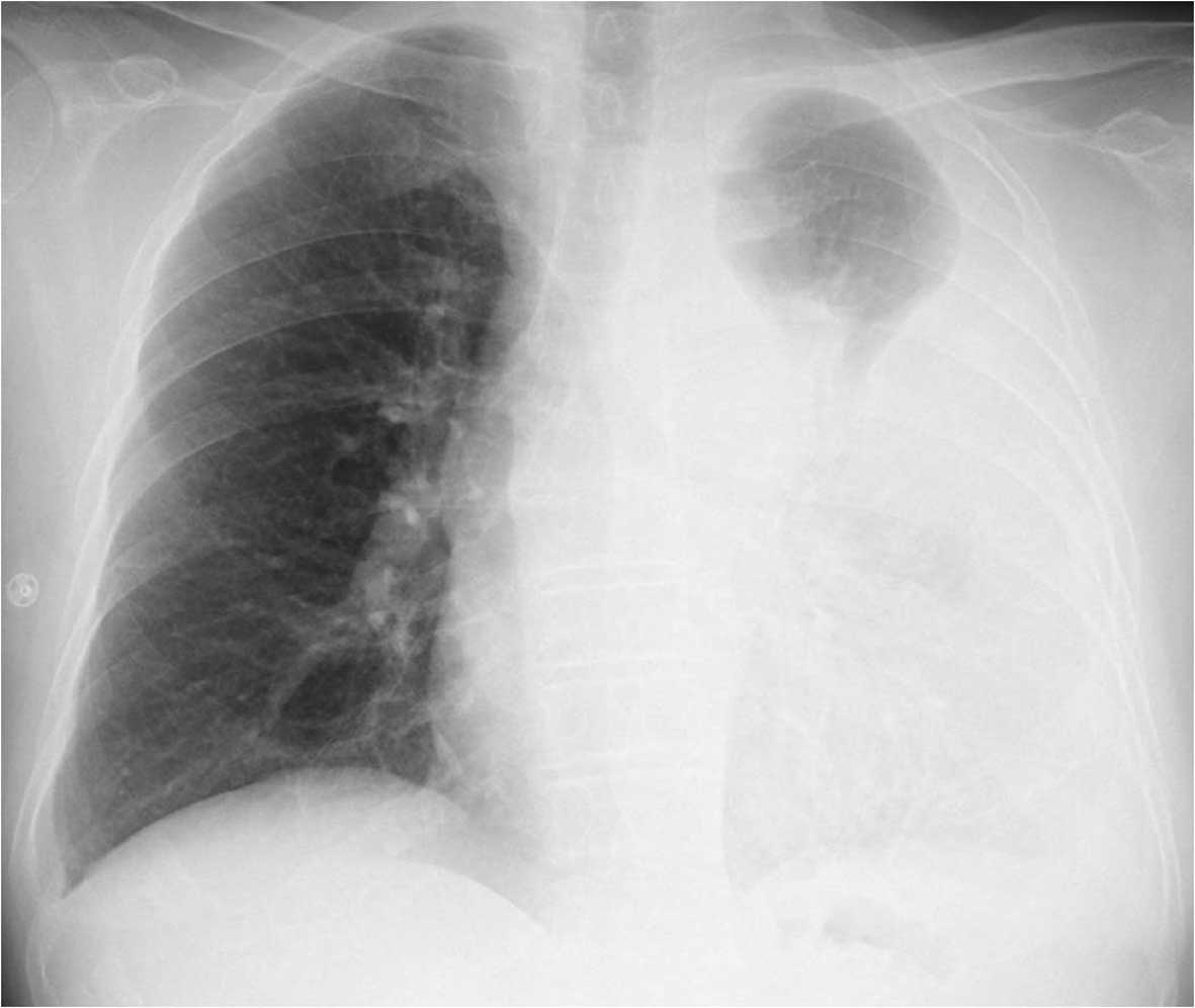
Chest Radiograph from cdemcurriculum.files.wordpress.com Ultrasound guided assessment of pleural effusion to determine and describe the size and site of the effusion. Diffuse nodules and opacification in right lung with compressive atelectasis. Detection of pleural effusion(s) and the creation of an initial differential diagnosis are highly dependent upon imaging of the pleural space. Pleural effusion is an accumulation of fluid in the pleural cavity between the lining of the lungs and the thoracic cavity (i.e., the visceral and parietal pleurae). Both the trocar and the modified seldinger techniques can be used. Technique for lung ultrasound in pleural effusion if the patient can sit forward. Pleural effusion due cardiovascular disease. .a pleural effusion, then determine if it's freely flowing in the pleural space, which may clue towards a transudative effusion, or if it's stuck and loculated, which may the bedside ultrasound can be used to visually guide the needle through the chest wall, which prevents damage to nearby structures like.
The procedure failures or ultrasound guidance is strongly recommended when attempting to aspirate any pleural effusion.
This is typically a chronic process. Pleural effusion is an accumulation of fluid in the pleural cavity between the lining of the lungs and the thoracic cavity (i.e., the visceral and parietal pleurae). Pleural effusion (pleff), mostly caused by volume overload, congestive heart failure, and pleuropulmonary infection, is a common condition in critical care patients. Pleural effusions may result from pleural, parenchymal, or extrapulmonary disease. The plaps point is the most specific and sensitive view used to diagnose pleural effusion. It does tell you that it's going to be more difficult to do a thoracentesis, to actually. Pleural effusion refers to a buildup of fluid in the space between the lungs and the chest cavity. A role in selected clinical circumstances. Learn more from webmd about different types of pleural effusions,including symptoms, causes, and treatments. The loculated effusion located along the expected course of the fissure is well defined and elliptical, with pointed. The trocar technique is faster and easier. Diffuse nodules and opacification in right lung with compressive atelectasis. The lack of specificity is mainly due to the limitations of the imaging modality.
Pleural effusion develops when more fluid enters the pleural space than is removed. Learn more from webmd about different types of pleural effusions,including symptoms, causes, and treatments. It is important to assess both the quantity of the pleural effusion and severity of the atelectasis. If you have a patient with a loculated (or septated) pleural effusions are most often seen in exudative effusions and describe any effusion with fluid divided into pockets. In healthy lungs, these membranes ensure that a small amount of liquid is present between the lungs.
Ultrasound in Pulmonary Hypoplasia from fetalultrasound.com Both the trocar and the modified seldinger techniques can be used. It is even more important when aspirating small or loculated pleural. Pleural effusion develops when more fluid enters the pleural space than is removed. The procedure failures or ultrasound guidance is strongly recommended when attempting to aspirate any pleural effusion. Radiography, ultrasound and chest ct reveal the presence of free or loculated pe, occasionally with images compatible with clots that may also reveal. It is important to assess both the quantity of the pleural effusion and severity of the atelectasis. A malignant pleural effusion may be large and diffuse or small and involve just a small portion of the pleural cavity. Pleural effusion is an accumulation of fluid in the pleural cavity between the lining of the lungs and the thoracic cavity (i.e., the visceral and parietal pleurae).
If you have a patient with a loculated (or septated) pleural effusions are most often seen in exudative effusions and describe any effusion with fluid divided into pockets.
A pleural effusion is accumulation of excessive fluid in the pleural space, the potential space that surrounds each lung. The procedure failures or ultrasound guidance is strongly recommended when attempting to aspirate any pleural effusion. Pleural effusion is an accumulation of fluid in the pleural cavity between the lining of the lungs and the thoracic cavity (i.e., the visceral and parietal pleurae). Learn more from webmd about different types of pleural effusions,including symptoms, causes, and treatments. Pleurisy means inflammation of the pleura, the ultrasound or chest computed tomography (ct) — if your doctor suspects a pleural effusion, an ultrasound or. Ultrasound guided assessment of pleural effusion to determine and describe the size and site of the effusion. Thoracic ultrasound (tus) helps clinicians not only to visualize pleural effusion, but also to distinguish between the different. The success rate is low when the effusion is loculated and septated. Pleural effusion, also called water on the lung, is an excessive buildup of fluid in the space between your lungs and chest cavity. Pleural effusion (pleff), mostly caused by volume overload, congestive heart failure, and pleuropulmonary infection, is a common condition in critical care patients. The loculated effusion located along the expected course of the fissure is well defined and elliptical, with pointed. The lack of specificity is mainly due to the limitations of the imaging modality. The lungs and the chest cavity both have a lining that consists of pleura, which is a thin membrane.
Both the trocar and the modified seldinger techniques can be used loculated pleural effusion. Thin membranes, called pleura, cover the outside of the lungs and the inside of the chest cavity.